Biomedical Research
A typical recording of the four physiological signals ©iStock.com/DNY59

Current technologies allow us to learn more about what is happening inside our bodies. Different modalities have been used in recent years to extract different information concerning our health: whether we measure the heart rate, blood pressure, blood glucose concentration, brain activity or use imaging modalities to get a better picture of our bones, eyes, muscles or nerves, we constantly generate information about our bodies that can help physicians diagnose certain diseases or aid at treating patients.

A biomedical signal can be any kind of signal that is measured from the human body, examples include: 1-D signals, when we measure vital signals, 2-D or 3-D images of the organs, or audio signals in hearing aids. Usually, the obtained biomedical measurements need to be analyzed in order for the clinician to be able to infer information from them. For example, in EEG signals, we commonly encounter motion artifacts that disrupt the signals and make it impossible to extract the necessary information. In Biomedical imaging, we often want to identify the different regions in an image using segmentation techniques and classify these to detect anomalies. Using methods of parameter estimation and models of the measurements, we can extract reliable information. Robust statistics play a big role in biomedical signal processing as real life data can often deviate from the model assumptions.

Many biomedical devices tend to be small or have to be affordable for the patient, which leads to very stringent conditions on power consumption and size of the devices. The challenge is often to design algorithms that comply with these constraints while showing high accuracy and reliability.

For more information on biomedical signal processing, see the sections below or contact the respective Research Associates.

Biolab

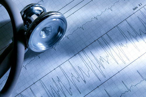
In our Biolab (Biological Experiment Laboratory), we have a selection of sensors to conduct measurements including electrocardiogram (ECG), photoplethysmogram (PPG), electrodermal activity (EDA), and respiration. For the simulation of activities, we provide a cross trainer and a treadmill. A project in cooperation with Roche Diagnostics GmbH, Mannheim, is on diabetes care; details can be found below.

See also SPG Lab for Body Worn Sensing of Physiological Parameters (opens in new tab) (Slides).

Projects

Our body constantly generates, senses, analyses and manipulates physiological signals. An emotional experience is associated with changes in physiological parameters, e.g., in the heart rate as shown in the figure, below. In cooperation with Prof. Dr. Augustin Kelava from Universität Tübingen, the Signal Processing Group investigates both the synchrony and the patterning of physiological response parameters. Together, we developed a new approach for the quantification of synchrony of multivariate non-stationary psychophysiological signals during emotion eliciting stimuli, which may allow the monitoring of mental wellbeing.

Physiological signals are non-stationary and artifacts affect measurements. Therefore, we developed advanced algorithms to robustly and accurately estimate physiological parameters, identify the synchrony between multiple synchronously measured signals, and classify and detect emotions.

For more information on this research project, or enquiries about Bachelor/Master thesis or Pro-/Project-Seminars, please contact Abdelhak Zoubir .

Change of hear rate (HR) for different participants of a psychological study.

Observing human gait plays a key role in medical diagnosis, biomedical engineering, physiotherapy and rehabilitation. For example, changes in gait patterns can reveal many neurological conditions, such as Parkinson’s, at an early stage.

Recently, radar has become of increased interest in many applications, including automotive, medical and consumer industries. Due to its non-intrusive, non-wearable and privacy-preserving sensing, it can serve as an effective tool for contact-less human gait monitoring. The back-scattered radar signal of a human walk contains multiple overlaying signal components from different parts of the body, e.g. arms and legs. Each of these components has its own Doppler shift depending on the velocity of the moving body part.

These so-called micro-motions lead to distinct micro-Doppler signatures, which are typically represented in the time-frequency domain. Based on micro-Doppler signatures different targets or motions can be discerned.

This project deals with radar-based human gait analysis. For this, we classify different human walking styles within the class of human gait. The idea is to use radar as a diagnostic tool, which can pave the way for ambulatory medical gait analysis.

For more information on this research project or enquiries about Bachelor/Master thesis or Pro-/Project-Seminars, please contact Abdelhak Zoubir

A person walking with a cane in front of a radar system.
Time-frequency representation of back-scatttered radar data from a walking person.

A common risk for patients with traumatic brain injuries is that the primary brain damage can lead to a secondary pathophysiological damage. To avoid such damages, the continuous monitoring of signals, such as ICP, mean arterial blood pressure (MAP) or brain tissue oxygen level (PtiO2), is essential.

This research project concerns the development of methods that describe the relationship between simultaneously observed signals. Since the signals are non-stationary and the measurements contain outliers and artifacts, advanced robust methods are required. We also develop methods for the robust forecasting of ICP signals. Accurate ICP forecasting enables active and early interventions for more effective control of ICP levels. To achieve high accuracy, most existing methods require a high sampling rate (100 Hz), which is infeasible for online medical applications. Therefore, we consider online ICP forecasting based a sampling rate 0.1 Hz.

For more information on this research project, or enquiries about Bachelor/Master thesis or Pro-/Project-Seminars, please contact please contact Abdelhak Zoubir .

ICP measurement with motion artifacts.

The human eye is a complex dynamic system consisting of several optical elements. It is well established that the eye’s wavefront aberrations fluctuate in time. Together with the Contact Lens and Visual Optics Laboratory (CLVOL), Queensland University of Technology (QUT), Brisbane, Australia, the Signal Processing Group conducted research to investigate the role of cardiopulmonary signals, i.e., pulse and respiration, in these fluctuations. We proposed a set of tools, based on joint time-frequency analysis to acquire a detailed picture.

We also developed statistical signal and image processing techniques in corneal modeling. Corneal topography estimation that is based on the Placido disk principle relies on good quality of pre-corneal tear film and sufficiently wide eyelid aperture to avoid reflections from eyelashes. However, in practice, these conditions are not always fulfilled resulting in missing regions, smaller corneal coverage, and subsequently poorer estimates of corneal topography. Our aim was to enhance the standard operating range of a Placido disk videokeratoscope to obtain reliable corneal topography estimates.

For more information on this research project, please contact please contact Abdelhak Zoubir .

Videokeratoscopic image with tear-film breakups and eyelashes shadows.
Reconstructed corneal topography.

Smartwatches or other wearable devices that contain optical heart rate monitors are an emerging technology that can be used, e.g as a tool to control the training load during physical exercises or to monitor physiologic conditions during daily activities. In contrast to the previous generation of devices, it is no longer necessary to wear an additional chest strap, since the heart rate is monitored from the wrist by means of photoplethysmography (PPG).

PPG refers to a noninvasive indirect measurement of the blood flow. Pulse oximeters illuminate the skin along with underlying blood vessels via light-emitting diodes (LEDs) to measure intensity changes of the reflected light that is absorbed by the photo diodes. Based on the intensity change in the PPG signal, the arterial oxygen saturation and the heart rate can be estimated. Combining the PPG signal with an electrocardiogram (ECG), allows the extraction of additional parameters, such as, e.g. the pulse arrival time (PAT) which is correlated with the blood pressure.

However, the PPG measurement is susceptible to motion artifacts, which inevitably occur during physical exercises. Motion induced artifacts can strongly deteriorate the quality of a PPG signal and signal processing techniques are sought for to remove the motion artifacts from the PPG signal prior to estimating the heart rate.

In this project, we develop new algorithms to robustly and accurately estimate the heart rate from PPG signals in the presence of motion artifacts. In order to apply our algorithms on real data, we also conduct measurement series and use external PPG data.

For more information on this research project, or enquiries about Bachelor/Master thesis or Pro-/Project-Seminars, please contact Abdelhak Zoubir .

Smartwatch with PPG sensor.
Measured PPG spectrogram.

Diabetes Mellitus is a chronic disease that affects 347 million people worldwide and results in high blood sugar levels. It can lead to severe medical complications, such as eye and nerve diseases. An essential component of diabetes care, which can help delay or even prevent complications is careful self-monitoring of blood sugar levels by the patients. To this end, invasive blood sugar measurement devices, as seen in the image on the right, are used up to 4-5 times daily by the patient. These devices need to show high accuracy and good usability, while remaining cheap under low power constraints.

In a cooperation project with Roche Diagnostics GmbH in Mannheim, we developed a new generation of blood sugar measurement devices that require blood sample volumes in the nl-range (100 times less than state-of-the-art devices). Hereby, the induced pain from extracting a blood sample from the finger is drastically reduced. A photometric measurement principle is used to extract the glucose concentration from the blood sample, where a camera observes the chemical reaction between the chemical test strip and the blood sample.

Using methods of statistical signal processing and image processing, we are able to segment the produced images to detect the region where the chemical reaction takes place and estimate the intensity value of the region. This is then mapped to the underlying glucose concentration. Furthermore, we aim at reducing measurement time to increase the convenience of measurement for the patient. We use state-space approaches combined with a model of the chemical kinetics to achieve this.

For more information on this research project, contact Abdelhak Zoubir .

A hand-held glucose monitoring device ©iStock.com/ratmaner
Fast glucose concentration estimation from a small blood sample.